
FRIDAY, July 29, 2022 (HealthDay Information) – The way forward for ultrasound imaging is usually a sticky label affixed to the surface that may transmit pictures regularly for 48 hours.
Researchers at Massachusetts Institute of Generation (MIT) have created a postage stamp-sized software that creates are living, high-resolution pictures. They reported on their growth this week.
“We imagine we have now opened a brand new technology of wearable imaging: With a couple of patches for your frame, that you must see your inner organs,” stated co-senior learn about writer Xuanhe Zhao, a professor of mechanical engineering and civil and environmental engineering at MIT.
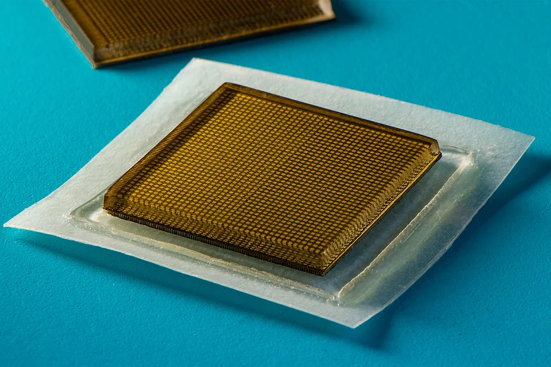
The sticky label — about 3/4-inch throughout and about 1/10-inch thick — is usually a exchange for cumbersome, specialised ultrasound apparatus to be had best in hospitals and physician’s place of job, the place technicians follow a gel to the surface after which use a wand or probe to direct sound waves into the frame.
The waves mirror again high-resolution pictures of a big blood vessels and deeper organs reminiscent of the center, lungs and abdomen. Whilst some hospitals have already got probes affixed to robot hands that can give imaging for prolonged sessions, the ultrasound gel dries through the years.
For now, the stickers would nonetheless must be hooked up to tools, however Zhao and different researchers are running on a method to perform them wirelessly.
That opens up the potential for sufferers dressed in them at house or purchasing them at a drug retailer. Even of their present design, they may get rid of the will for a technician to carry a probe in position for a very long time.
Within the learn about, the patches adhered smartly to the surface, enabling researchers to seize pictures even though volunteers moved from sitting to status, jogging and cycling.
“We envision a couple of patches adhered to other places at the frame, and the patches would keep in touch along with your cell phone, the place AI algorithms would analyze the photographs on call for,” Zhao defined in an MIT information free up.
A distinct manner examined — stretchable ultrasound probes — yielded pictures with deficient decision.
“[A] Wearable ultrasound imaging instrument would have large doable at some point of scientific prognosis. On the other hand, the decision and imaging length of current ultrasound patches is somewhat low, and so they can not symbol deep organs,” stated co-lead writer Chonghe Wang, a graduate scholar who works in Zhao’s Lab.
The MIT crew’s new ultrasound sticky label produces upper decision pictures through pairing a stretchy adhesive layer with a inflexible array of transducers (they convert power from one shape to some other). Within the heart is a cast hydrogel that transmits sound waves. The adhesive layer is constituted of two skinny layers of elastomer.
“The elastomer prevents dehydration of hydrogel,” co-lead writer Xiaoyu Chen defined. “Handiest when hydrogel is very hydrated can acoustic waves penetrate successfully and provides high-resolution imaging of inner organs.”
Wholesome volunteers wore the stickers on quite a lot of spaces, together with the neck, chest, stomach and hands. The stickers produced transparent pictures of underlying buildings, together with the converting diameter of primary blood vessels, for as much as 48 hours. They stayed hooked up whilst volunteers sat, stood, jogged, biked and lifted weights.
They confirmed how the center adjustments form because it exerts all over workout and the way the tummy swells, then shrinks, as volunteers drank after which eradicated juice. Researchers additionally may hit upon indicators of transient micro-damage in muscular tissues as volunteers lifted weights.
“With imaging, we may be able to seize the instant in a exercise sooner than overuse, and forestall sooner than muscular tissues develop into sore,” Chen stated. “We have no idea when that second may well be but, however now we will supply imaging knowledge that professionals can interpret.”
Along with running on wi-fi generation for the stickers, the crew is creating instrument algorithms in response to synthetic intelligence that may higher interpret the ultrasound pictures.
Zhao thinks sufferers would possibly someday be capable to purchase stickers that may be used to watch inner organs, the development of tumors and building of fetuses within the womb.
“We believe we may have a field of stickers, every designed to symbol a special location of the frame,” Zhao stated. “We imagine this represents a leap forward in wearable units and clinical imaging.”
The findings had been revealed July 28 in Science.
Additional info
The Nationwide Institute of Biomedical imaging and Bioengineering has extra on ultrasound.
SOURCE: Massachusetts Institute of Generation, information free up, July 28, 2022
Via Cara Murez HealthDay Reporter

Copyright © 2021 HealthDay. All rights reserved.

SLIDESHOW
Well being Screening Exams Each and every Girl Wishes
See Slideshow